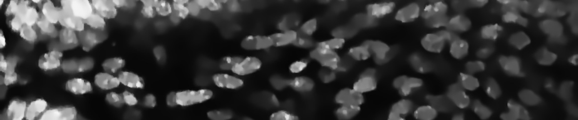
Automatic quantification of immunofluorescence images relies either on the detection and counting of spots superimposed on biological structures, usually immersed in a non-uniform background, or on the outlining of larger cellular compartments. We have developed methods for spot detection and characterization that allow a fast and reproducible quantitative analysis of these images. based on a multiscale approach that uses a shift invariant discrete wavelet transform(SI-DWT) and on the selective filtering of wavelet coefficients. This scheme allows to separate and characterize objects of different sizes by selecting only a vicinity of detail images with corresponding scales adapted to the size of the spots. The extraction step consists in retaining the significant responses of the locally supported detail signal filters to the desired features, at the different scales of the wavelet representation. This is accomplished through a denoising technique using a threshold value which is image and level dependent and which can be computed automatically from the data. The program detects spots both in 2D and in 3D images and is used by several biological scientists to quantify their images.
Spot analysis in immunofluorescence images
September 21, 2011 | 0 comments