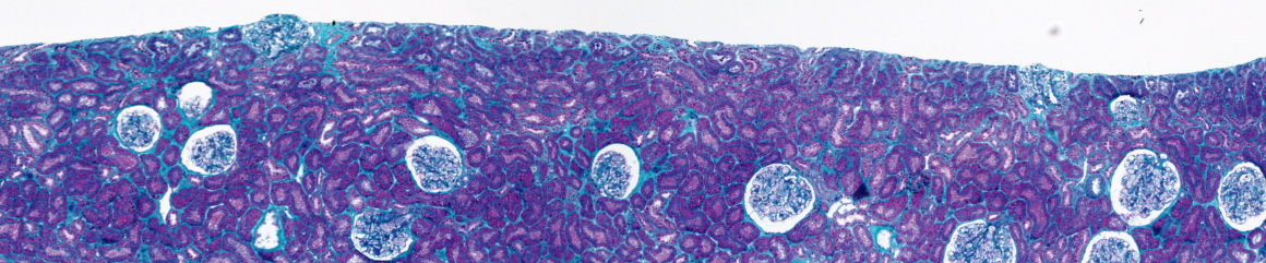
To characterize histopathological images stained by different colorations, we have developed a system to analyse color images. Images are segmented by a split and merge approach and by color quantization, to reduce color classes. We have also defined a criterion to choose the best color space. This method has been applied successfully to the quantification of interstitial fibrosis quantification in chronic allograft nephropathy of renal biopsy. The proportion of green to total pixels in the biopsy was then calculated and used as an index of interstitial fibrosis. The results are correlated to the values of quantification realized by an expert.
Color image analysis in histology
September 21, 2011 | 0 comments