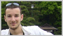
Dr. Alexandre Dufour graduated in Artificial Intelligence & Pattern Recognition from University Pierre & Marie Curie (Paris, France) in 2004, and performed his doctoral research at Institut Pasteur Korea (Seoul, South Korea) from 2005 to 2007, where he co-founded the Image Mining group as a Junior Scientist. He then pursued his postdoctoral work in the Quantitative Image Analysis group at Institut Pasteur (Paris, France), and is now a permanent Staff Scientist in the group, leading the « cell deformation and motility » research team.
Dr. Alexandre Dufour is a member of the Institute of Electrical and Electronics Engineers (IEEE), the IEEE Signal Processing Society, an associate member of the IEEE Bio-Imaging and Signal Processing Technical Committee, and also a board member of the “Functional Live Microscopy” development group (GdR 2588) of the French National Council for Scientific Research (CNRS). His teaching activities include the EMBO course « Imaging Parasite Infection » and the CNRS school on live microscopy « MiFoBio ».
Research
ANALYSIS AND MODELING OF CELL MORPHODYNAMICS
Elucidating the mechanisms of cell deformation and motility is a topic of major interest in cell biology, for they are strongly involved in cell development, immune responses, cancer and infectious diseases. Our aim to develop robust and fully automated image analysis tools, able to extract quantitative measures of cell dynamics from multi-dimensional (2D/3D), multi-modal (brightfield/phase-contrast/fluorescence) time-lapse microscopy data. It is nowadays a growing consensus that the combination of large-scale cell imaging and sophisticated computational analysis tools is necessary to provide new insights on numerous biological phenomena. Yet, many studies related to cell shape and motility rely still today on manual (though generally computer-assisted) analysis in 2D sequences. This process is cumbersome and prone to user bias and reproducibility issues. Migration to full 3D time-lapse experiments is a necessary step to reliably quantify cell shape information, but induces new challenges in terms of image acquisition and quantification, yielding a need for more robust and fully automated analysis techniques.
For many years now we have been developing automated cell tracking approaches based on the well-known formalism of deformable models, a.k.a. “active contours”. The principle is to deform an initial contour placed on the image until it fits the boundary of the target cell. The deformation can be mathematically expressed as the minimization of an energy functional, which comprises several terms related either to the image data (driving the contour toward the cell boundary) or to geometrical properties of the contour (regularizing the deformation to avoid local energy minima). This energy is an essential ingredient of the method, and several of our contributions consist in adapting this energy and its implementation to meet the particular constraints of biological imaging, e.g., low signal-to-noise ratio, multiple cell-cell contacts over time, inhomogeneous cell staining, photo-bleaching, etc.. Active contours also offer a flexible formalism for efficient shape representation and quantification, allowing to compute robust statistics over large-scale cell populations.
SOFTWARE DEVELOPMENT FOR EXTENDED REPRODUCIBLE RESEARCH
Over the past decade, image analysis and quantification in biology has become an established research field, now going by the name of “BioImage Informatics”. The tools developed in this field are dedicated to understanding biology through proper, systematic quantification by means of imaging, yielding issues of big data management, automated quantification, and reproducibility. Our group has founded and leads the development of the Icy software, an open-source platform for reproducible research in bioimaging. Icy is community-oriented and brings together the biology, imaging and computer vision communities by providing an integrated software + web site that provides (among other features): 1) cutting edge image processing and 2D/3D visualization algorithms to the end-user; 2) a simple developer toolkit for fast development (notably elegant graphical interfaces); 3) a content management system online for simple delivery of tools and automatic updates; 4) a graphical interface letting end-users tailor their own custom analysis in the form of reproducible protocols, without any knowledge about computer programming; 5) compatibility with existing software packages including ImageJ, Matlab & R but also programming languages such as Javascript and Python. We strongly believe that developing efficient analysis algorithms cannot be achieved without proper associated software tools to disseminate said algorithms to the community, and that such tools should benefit to both users and developers, every step of the way. Icy already has a well-established, and is being used in many major research groups and bioimaging platforms all over the world. See more at http://icy.bioimageanalysis.org.
Selected Publications
(full list available at Google Scholar or ResearchGate)
- Alexandre Dufour, Tzu-Yu Liu, Christel Ducroz, Robin Tournemenne, Beryl Cummings, Roman Thibeaux, Nancy Guillen, Alfred Hero, Jean-Christophe Olivo-Marin, “Signal Processing Challenges in Quantitative 3-D Cell Morphology: More than meets the eye“. IEEE Signal Processing Magazine 32 (January 2015). http://ieeexplore.ieee.org/xpls/abs_all.jsp?arnumber=6975298
- Sorin Pop*, Alexandre Dufour*, Jean-François Le Garrec, Chiara Ragni, Clémire Cimper, Sigolène Meilhac, Jean-Christophe Olivo-Marin, “Extracting 3D cell parameters from dense tissue environments: application to the development of the mouse heart“. Bioinformatics 29 (June 2013) pp. 772-779. http://www.ncbi.nlm.nih.gov/pubmed/23337749
- Fabrice de Chaumont, Stéphane Dallongeville, Nicolas Chenouard, Nicolas Hervé, Sorin Pop, Thomas Provoost, Vannary Meas-Yedid, Praveen Pankajakshan, Timothée Lecomte, Yoann Le Montagner, Thibault Lagache, Alexandre Dufour, Jean-Christophe Olivo-Marin, “Icy: an open bioimage informatics platform for extended reproducible research“. Nature Methods 9 (July 2012) pp. 690-696. http://www.ncbi.nlm.nih.gov/pubmed/22743774
- Roman Thibeaux, Alexandre Dufour, Pascal Roux, Michèle Bernier, Anne-Catherine Baglin, Pascal Frileux, Jean Chrisophe Olivo-Marin, Nancy Guillén, Elisabeth Labruyère, “Newly visualized fibrillar collagen scaffolds dictate Entamoeba histolytica invasion route in the human colon“. Cellular Microbiology 14 (February 2012) pp. 609-621. http://www.ncbi.nlm.nih.gov/pubmed/22233454
- Alexandre Dufour, Roman Thibeaux, Elisabeth Labruyère, Nancy Guillén, Jean-Christophe Olivo-Marin, “3-D active meshes: fast discrete deformable models for cell tracking in 3-D time-lapse microscopy“. IEEE Transactions on Image Processing 20 (September 2011) pp. 1925-1937. http://www.ncbi.nlm.nih.gov/pubmed/21193379
- Alexandre Dufour, Vasily Shinin, Sharagim Tajbakhsh, Nancy Guillén, Jean-Christophe Olivo-Marin, Christophe Zimmer, “Segmenting and tracking fluorescent cells in dynamic 3-D microscopy with coupled active surfaces“. IEEE Transactions on Image Processing 14 (September 2005) pp.1396-1410. http://www.ncbi.nlm.nih.gov/pubmed/16190474
*: equal author contribution
Contact
Phone: + 33 1 45 68 83 92
Email: adufour@pasteur.fr


